docplexus
Case Presentation A 26-year-old G1P0 woman with a history of systemic lupus erythematosus, Sjögren’s syndrome, and scleroderma presented for high-risk delivery. She was transferred to the facility because of hypoxia requiring up to 10 L of supplemental oxygen and fetal bradycardia with prolonged decelerations. On arrival, she was found to have severe preeclampsia and underwent emergency caesarean delivery.
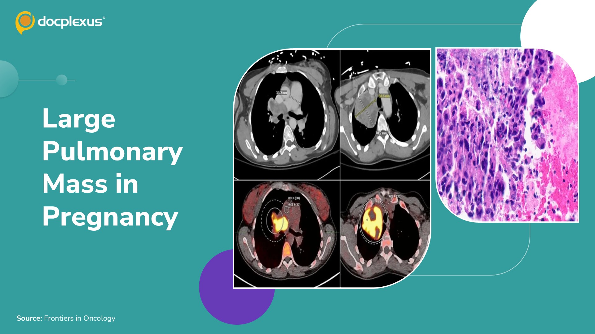
Recent medical history Three-month history of persistent cough Sought care at another hospital Current presentation Chest computed tomography (Figure 1) 12-cm mediastinal mass invading the right pulmonary artery with near-complete occlusion Encirclement of the right mainstem bronchus Figure 1. CT and PET-CT demonstrating the large mediastinal lesion and pulmonary artery involvement. C-section delivery Underwent emergency caesarean delivery at 36 weeks and 6 days Gave birth to a…