
Growth hormone deficiency (GHD) stems from dysfunction in the pituitary gland or disruptions in the hypothalamic–pituitary axis that controls GH regulation. 1 Patients with GHD exhibit significantly smaller volumes in brain structures such as the globus pallidum, thalamus, splenium of the corpus callosum, and hippocampus compared to those with idiopathic short stature. Imaging techniques like structural MRI, surface-based morphometry (SBM), voxel-based morphometry (VBM), and diffusion tensor imaging (DTI) are valuable tools for assessing structural and microstructural brain changes in GHD.
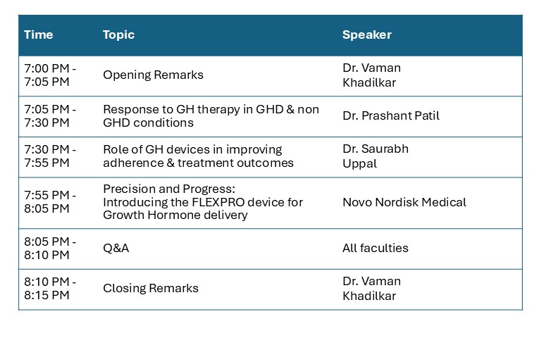
2 To improve treatment adherence, advancements have been made in the design of growth hormone delivery devices and the development of long-acting formulations. One such innovation is FlexPro®, a user-friendly, preloaded, premixed, and disposable multidose pen that requires minimal force for injection…