docplexus

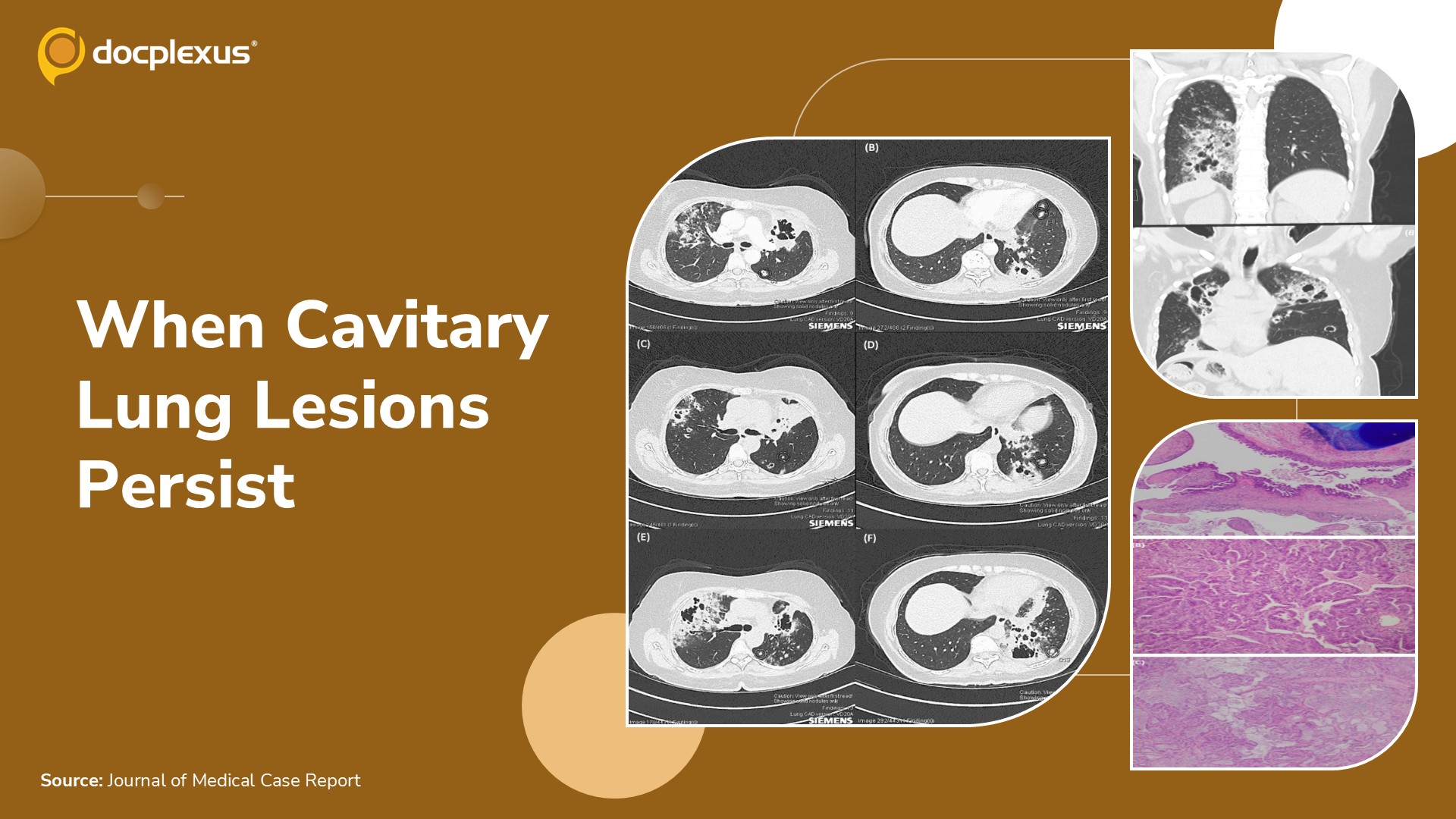
A 59-year-old woman presented with worsening cough and fever despite oral antibiotic therapy. She was referred by her general practitioner for inpatient management of presumed pneumonia. Her medical history included type 2 diabetes mellitus and hypertension.
She had never smoked and had no family history of cancer. Initial laboratory investigations C-reactive protein (CRP): 840 mg/L Erythrocyte sedimentation rate (ESR): 85 mm/hour White blood cell count: Within normal limits Interferon-gamma release assay: Negative Serum Aspergillus antigen: Negative Autoantibody screening: Negative Initial chest computed tomography (CT) (Figure 1A and 1B) Patchy inflammatory changes in the right upper lobe Large cavitary lesion in the left upper lobe Smaller cavitary lesion in the left lower lobe Extensive pulmonary infiltrates in the upper lobes Multiple small areas of cavitation bilaterally, with…