
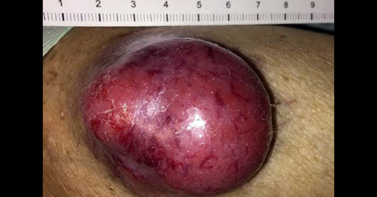
A woman in her 60s presented with multiple painful cutaneous nodules that had developed over the previous 2 months. History The patient had a history of IgG k multiple myeloma, diagnosed approximately 6 months earlier. At that time, flow cytometry showed a k-restricted plasma cell population positive for CD38 andCD138 but negative for CD3, CD20, and CD45 expression. Cytogenetic analysis with fluorescent in situ hybridization did not reveal high-risk genetic markers.
There was no evidence of end organ disease and her myeloma was classified as stage II. Since her initial diagnosis, she had completed 4 cycles of lenalidomide, bortezomib, and dexamethasone (RVD) therapy in addition to irradiation for lytic lesions of the thoracic and lumbar spine. Physical examination Scattered red-violaceous nodules with the largest measuring approximately 6.0×5.5×2.5cm (Image attached). They were found in…