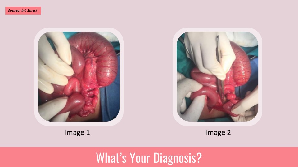
Recently we operated a case of an ectopic testis. The testis was reported in ultrasound and MRI to be present in the right iliac fossa. The region was explored and structure fitting into the described structure, apparently looking like atrophic testis, was excised and sent for biopsy. Biopsy reported it as fibro-fatty tissue containing lymphoid tissue.
Repeat ultrasound now reported the testis at a point just deep to the anterior abdominal wall, 9 centimetres lateral to umbilicus 2.5 centimetre medial to the spino-umbilical line. When I examined this patient on ultrasound, I found this tissue be hypoechoic and almost similar to a lymph node. Since the location was not in line with testicular descent, I preferred to have the opinion of a specialist ultrasonologist, who labelled this tissue to be ectopic testis. Now the question is what is echogenicity of the ectopic testis, how does it…