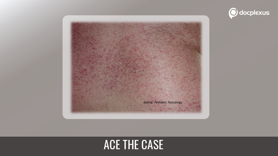
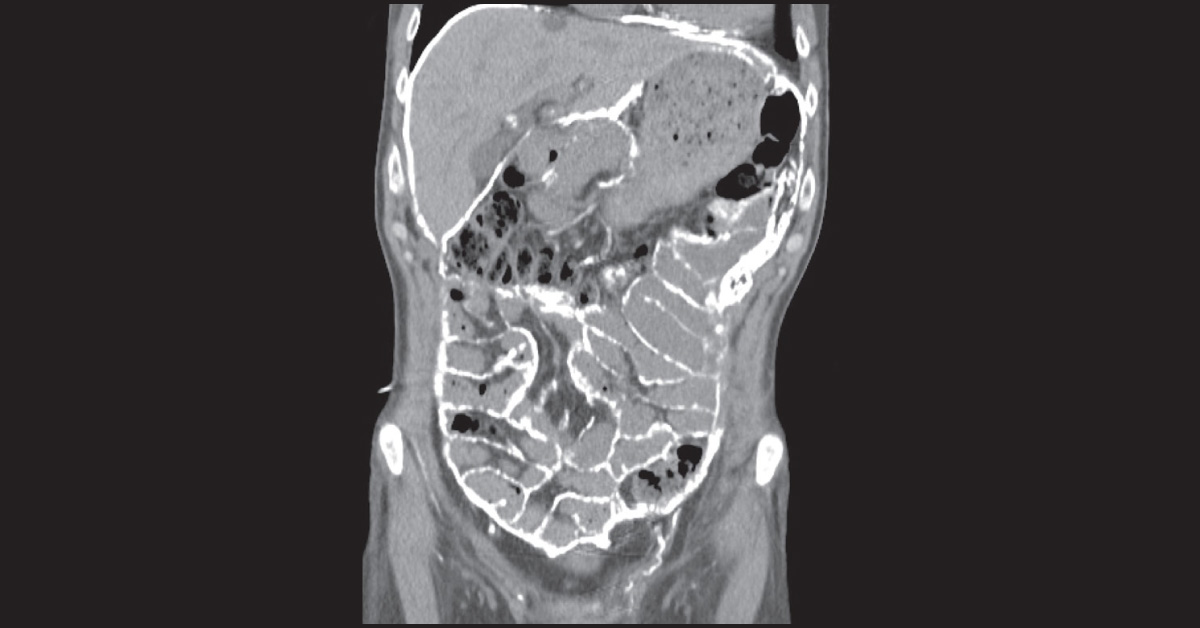
A 45-year-old man with end-stage renal failure secondary to Alport syndrome was presented with abdominal pain and hemodynamic instability. He has been on the maintenance peritoneal dialysis for 19 years. Examination & Procedure His abdomen was tender and 500Ml of fresh blood was drained from the peritoneal dialysis catheter. Following this, abdominal CT scan was performed. CT scan showed thickened bowel wall and calcification of abdominal viscera (first two images) At laparotomy, exploration of the abdomen showed severe calcification (last image) of the abdominal wall and bowel loops.
Blood was seen arising from the pelvis, which was subsequently packed with gauze, stopping the hemorrhage. However, in your opinion, what is the most likely reason of calcified abdomen? *This case is from Docplexus editorial team for education purpose only. Source: The Lancet