
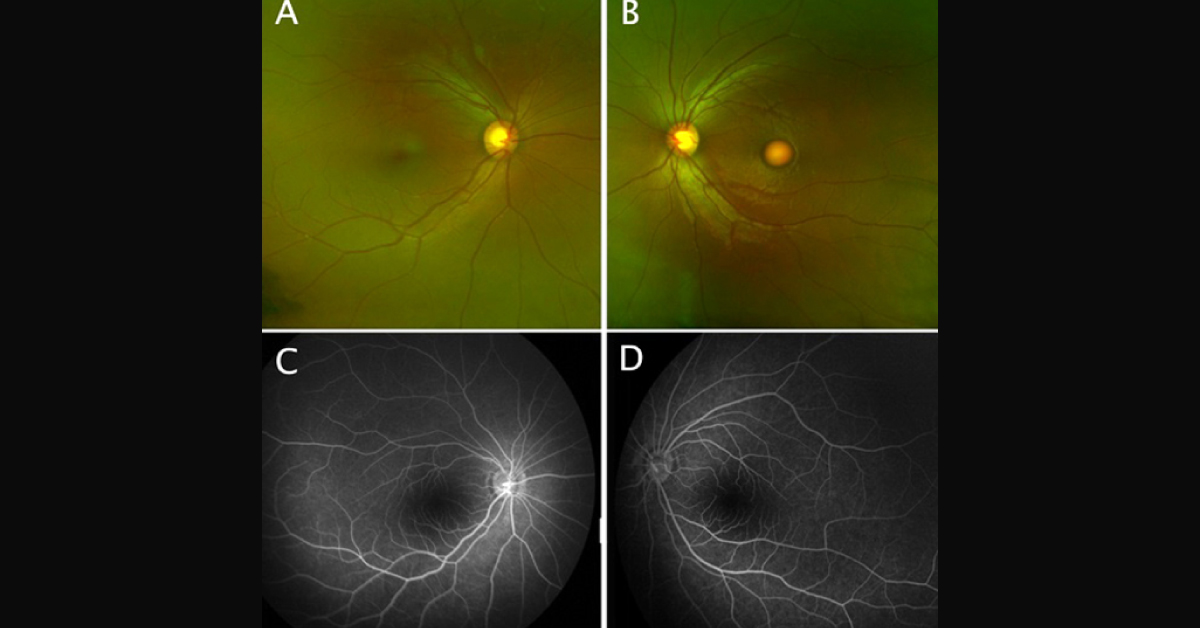
A 5-year-old girl was presented with decreased vision in her left eye. She was born full term by normal vaginal delivery. Past ocular, medical, and family histories were unremarkable. Her parents reported no ocular trauma. She was not on medications and immunization records were up-to-date. Examination Right visual acuity was 20/25 and 20/30 for the left eye. Ocular motility examination was normal.
No apparent pupillary effect was found. Both lenses were clear IOP was 19 mmHg in the right eye and 20 mmHg in the left eye. Fundus examination revealed a normal macular and vitelliform macular lesion in the left eye (Image A,B). Late-stage fundus fluorescein angiogram (C,D) and infrared reflectance imaging with corresponding SD-OCT foveal cross section (E,F) of both eyes demonstrated asymmetric disease. The optic nerve heads and peripheral retinae of both eyes appeared normal. What is the…