
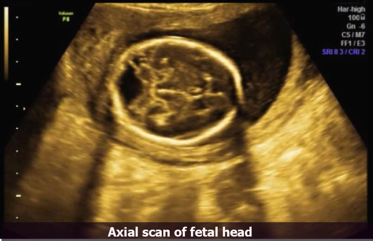
A 34-year-old woman who was 22 weeks pregnant, underwent a routine transabdominal obstetric ultrasound examination to exclude any congenital fetal abnormalities. Ultrasound scan findings - There is a triangular defect towards the posterior aspect or occipital region of the fetal brain. The mid-sagittal sections of the fetal brain, show the triangular defect posterior to the cerebellum.
There is an enlarged posterior fossa, cystic dilatation of the fourth ventricle, no significant dilatation of the lateral ventricles, near absence or hypoplasia of the cerebellum and the vermis, and elevation of the tentorium cerebelli. Clinical examination - Vital signs are normal, BP 124/82 mm Hg, abdominal examination showed gestational age of 22 weeks PMH - Before pregnancy menses were regular, she has no vaginal bleeding for 22 weeks Fig 1- Axial scan of the fetal head Fig 2- The second view of an…





