
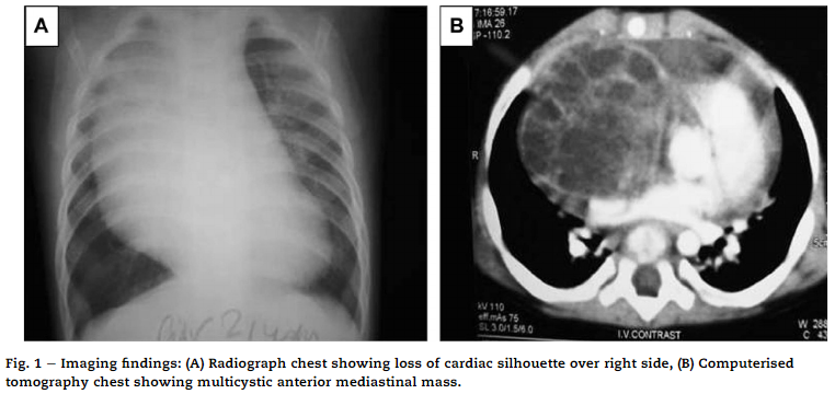
A 6-month-old male patient was brought to pediatric outpatient with complaints of respiratory distress. On general physical examination, the child was afebrile and active with a regular heart rate of 138/min. There was no pallor, icterus, cyanosis, or lymphadenopathy present. Systemic examination was within normal limits. All the hematological and biochemical parameters were also within normal limits. The radiograph chest showed a homogeneous opacity on the right side of the chest with smooth convex lateral margins.
Medial margins could not be seen and there was a loss of cardiac silhouette over the right side (Fig. 1A). Echocardiography showed a visible mass abutting the cardiac chambers. Computerized Tomography (CT) thorax revealed: multi-cystic anterior mediastinal mass, measuring 6*6*4cm, which was well-circumscribed, multiloculated, and intra-pericardial in location. It was…