
Case Presentation A 29-year-old pregnant woman was referred for evaluation of male fetal megacystis and bilateral ureterohydronephrosis identified on ultrasound (US) at the 20 th week of gestation. The subsequent US showed persistent bladder and ureteric dilations as well as pelvicaliectasis. The amniotic fluid volume was adequate and kept stable. A cesarean section was performed at 39th gestational week.
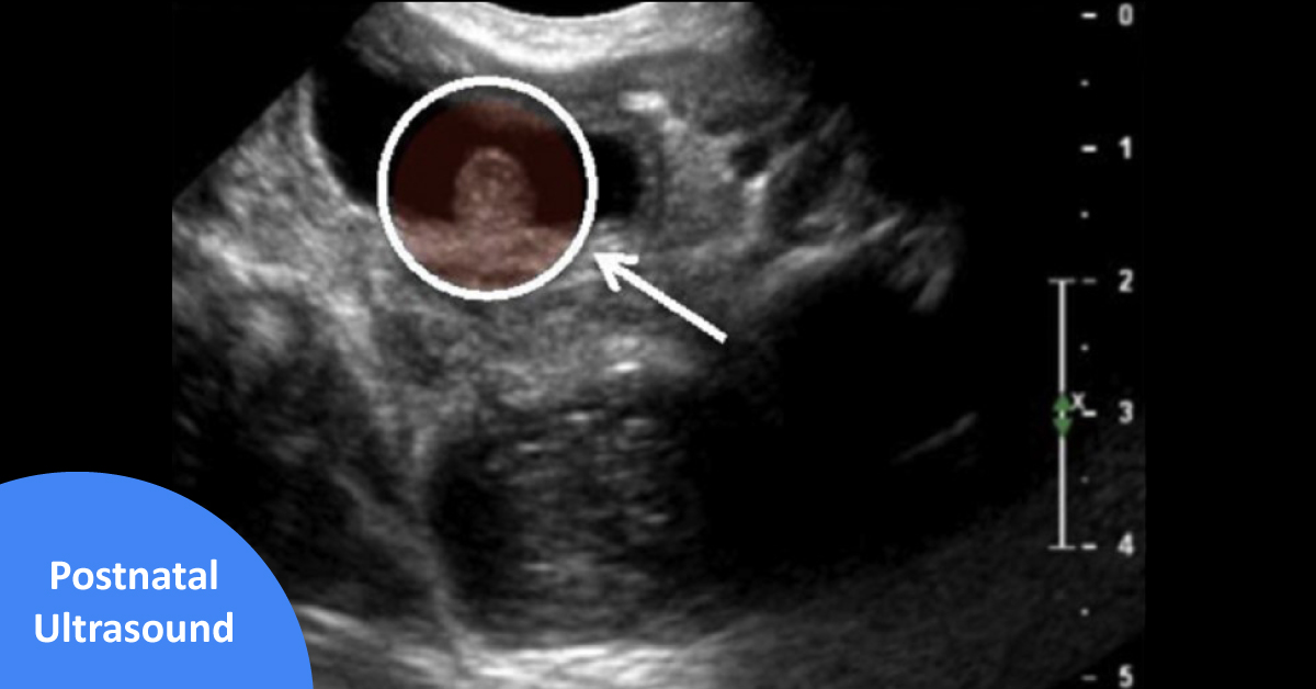
Neonatal physical examination showed an abnormally distended abdomen with a palpable bladder; the abdominal musculature was normal and no other abnormal features were identified. The bladder was easily catheterized with immediate drainage of clear urine. Postnatal US showed bilateral grade V ureterohydronephrosis (SFU) and an enlarged thick-walled bladder; an echogenic round pediculated mass arising from the proximal urethra was also identified (Image 1). The voiding cystourethrogram…