
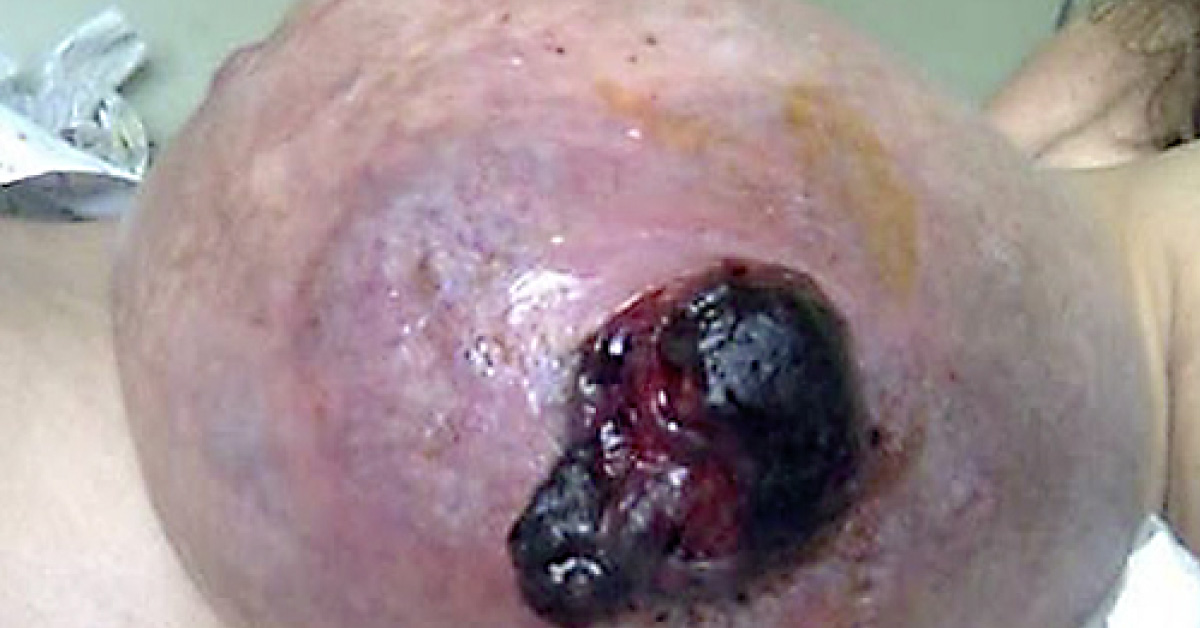
Case A 52-year-old woman with a normal mammography 2 years before, was presented an extremely voluminous tumor in the left breast which appeared 10 days before admission. A few hours after admission the breast started to ulcerate and bleed. Physical examination revealed a very large tumor with (>20 cm) bosselated contours, was hypervascularized, firm, and had a hemorrhagic area. Ulceration increased dramatically in 3 days and hemoglobin dropped to 8.8 g/dl Core biopsy indicated malignant phyllodes tumor, with areas of primary sarcoma.
Breast MRI revealed no muscular invasion and staging for systemic metastases was completely negative. A total mastectomy was performed and histopathology indicated a malignant phyllodes tumor, with 24 cm of longer axis, widely ulcerated, and the malignant mesenchymal component matched a high-grade leiomyosarcoma. The sentinel lymph node was free of…





