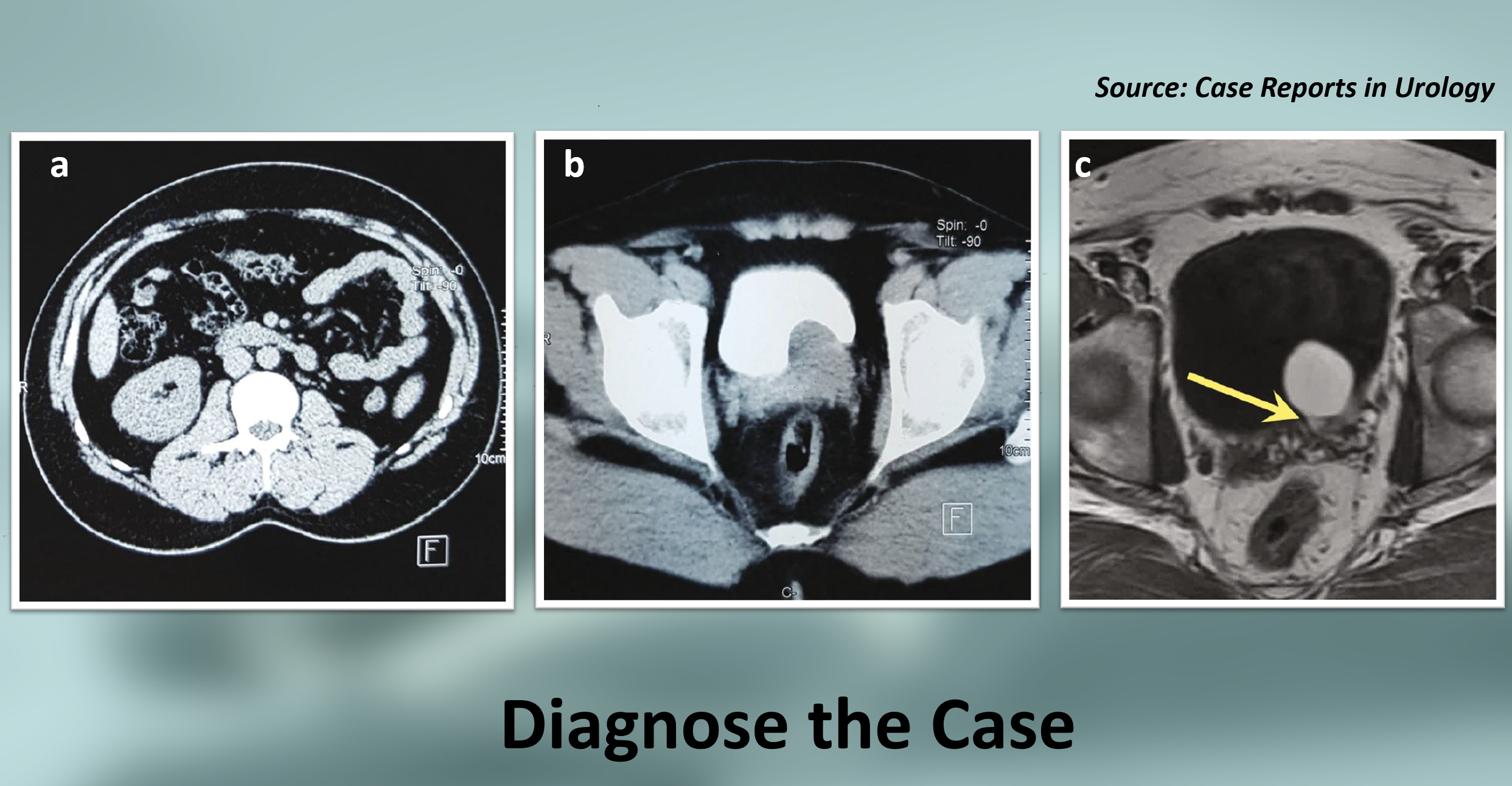
Case presentation A 33-year-old male patient reported to the urology department with apyretic recurrent dysuria, along with hematospermia episodes and painful ejaculation for the past 5 years. No medical or surgical history was reported by the patient. Radiographic findings Abdominal and pelvic computed tomography (CT) showed an empty left renal fossa (Figure a). A large left retrovesical mass was seen compressing the urinary bladder, suggestive of a cystic mass (Figure b). Pelvic magnetic resonance imaging (MRI) showed a unique cystic-to-tubular structure in the seminal vesicle.
The yellow arrow shows the tubular structure linked to the seminal vesicle cyst (Figure c). Cystic-to-tubular structure size: 20 × 35 × 40mm. Other investigations Digital rectal examination (DRE)- a renitent mass was found facing the rectum’s anterior wall. Blood and urine laboratory tests- no abnormalities…