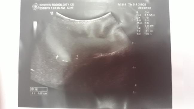
Dear Colleagues, Please help. I have a 15-year-old daughter with no significant past medical history nor trauma. She complains of LUQ pain on and off. An abdominal US Scan showed a single, nearly rounded, solid hypoechoic splenic focal lesion, 1.6 cm in diameter, in the mid-zone near the hilum. What is astonishing is that this lesion did not appear in MRI with diffusion. There is no fever nor lymphadenopathy. The longest splenic diameter on the US is 11.7 cm. Her CBC only showed monocytosis 8% with normal TLC and platelets while her Hb was 11.7 normocytic normochromic. LDH, CRP, renal function was all normal.
BRUCELLA, CMV, EBV Serology was all negative. Thrombophilia screen was unremarkable. What showed I do? All the story happened in the last 4 weeks. please help. What is the Differential Diagnosis? What is next? Follow up, or Aspirate, or computed tomography with Contrast, or…