
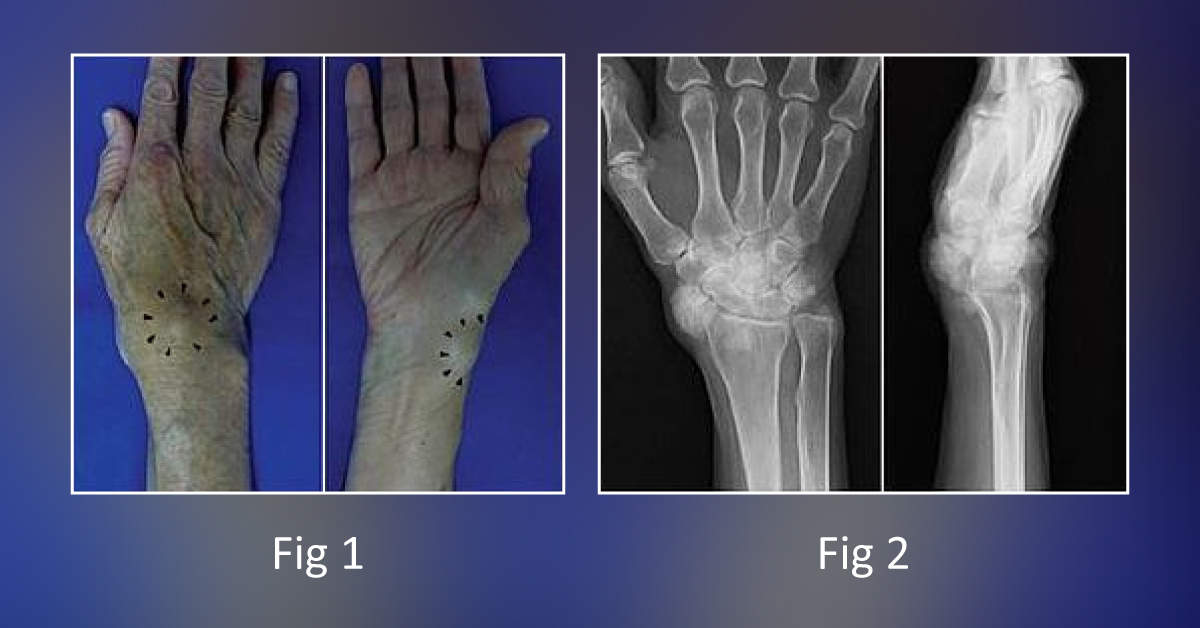
Case Presentation: A 78-year-old woman having a 5-year history of nodular lesions on the right wrist was presented to the hospital ward. Patient had no previous history of gout, hyperparathyroidism, hemochromatosis, or hypothyroidism. Physical Examination: Radial nodular lesion: (2 × 2) cm Dorsal nodular lesion: (2 × 2.5) cm Nodular lesions were non-movable, painful, or tender [Figure 1] Radiological Findings: Right wrist joint X-Ray: Multiple periarticular nodular lesions with calcifications (lesion was not palpable on the skin).
[Figure 2] Computed tomography (CT): Multiple calcified nodular lesion. [Figure 3] Magnetic resonance imaging (MRI): Nodular lesions around the right wrist joint showed low signal intensity on T1-weighted images and a mixture of high- and isosignal intensity on T2-weighted images. [Figure 4 (a, b)] Laboratory studies such as serum uric acid, calcium,…