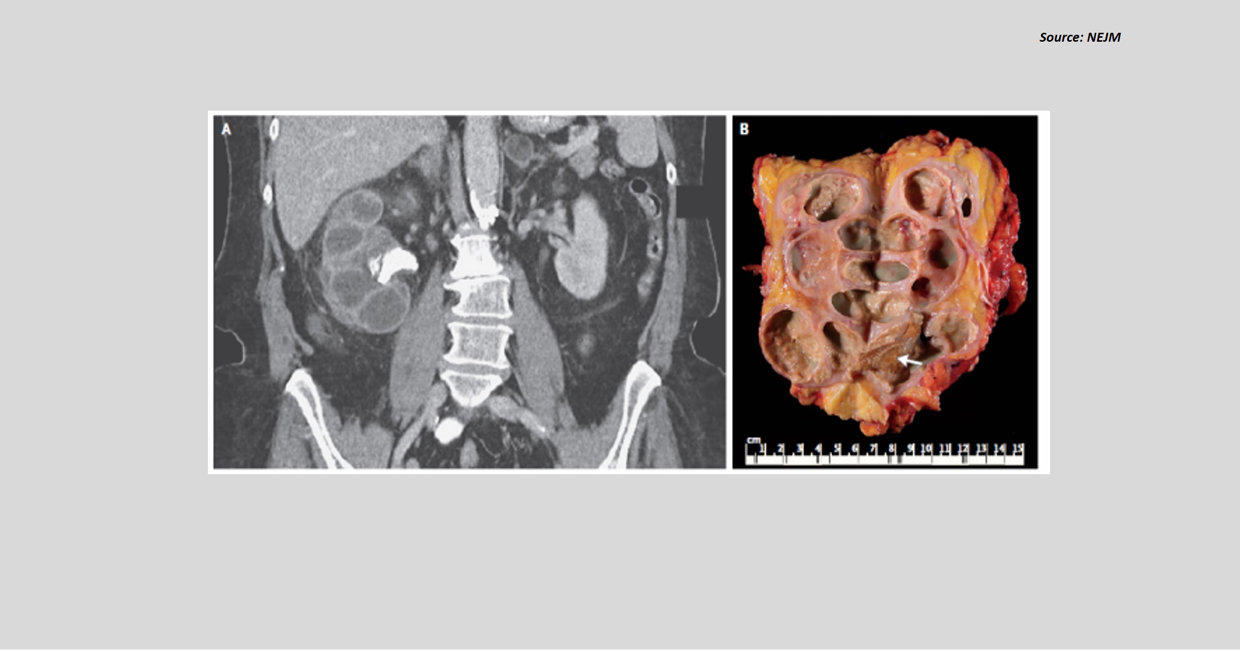
Case presentation A 72-year-old female patient presented with a 10-day history of fever and lethargy. The patient reported a history of type 2 diabetes mellitus, recurrent urinary tract infection, and weight loss of approximately 13.5 kg. Laboratory findings WBC count: 14,290/ μL Serum Creatinine: 1.3 mg/dL Urine Culture: Negative for bacteria Other investigations CT of the abdomen showed an enlarged right kidney with staghorn calculus in the ureteropelvic junction, cortical atrophy, and dilatation of calyces (Figure A). Percutaneous nephrostomy was performed, and culture from drainage yielded Proteus mirabilis.
Right side nephrectomy was performed as the radionucleotide scan showed the absence of renal function of the right kidney. Gross pathological analysis showed a staghorn calculus, dilated calyces, cystic cavities, and yellow tissue (Figure B). Based on the classical CT image and…