
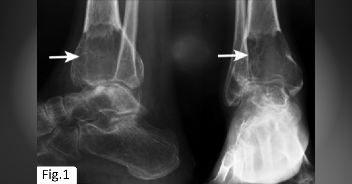
Case presentation A 42-year-old female was presented to the Orthopedic OPD with complaints of pain and swelling in the right leg from the last six months. The patient had no history of trauma. Clinical examination 1. Right ankle skiagram showed an expansile lytic lesion in epimetaphysis of the lower end of the tibia, causing pencil thinning of the adjacent cortex with a narrow zone of transition. 2. Soap bubble appearance due to multiple septations in the lesions. 3.
Serum calcium level 8.5 mg/dl, serum phosphorous 4.4 mg/dl, alkaline phosphatase 82IU/L, and C-reactive protein level was normal. 4. Radiology diagnosis showed osteoclastoma (Figure 1), and biopsy revealed a giant cell lesion at another center. 5. Microscopic study revealed a tumor made of fibrofatty tissue showing large areas of osteoid and bone trabeculae (Figure 2). The findings did not correlate with conditions such as…





