
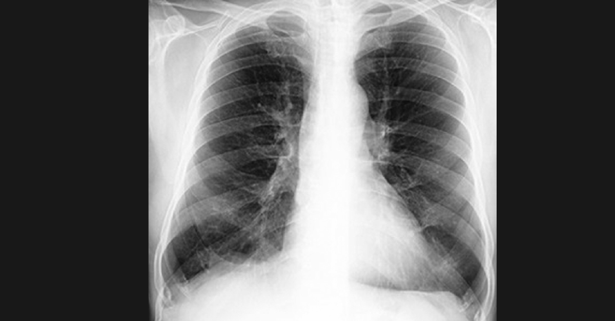
Case A 63-year-old nonsmoker man was presented with 10-year complaints of recurrent dry cough and progressive exertional dysponea. No history of hemoptysis, joint pains, fever, chest pain, or any constitutional symptoms. Past and family histories were unremarkable Examination Chest X-ray showed hyperinflated lung field with low flat diaphragms (figure attached). The ECG showed a poor progression of R-waves.
A high-resolution CT scan of the chest showed bilateral centrilobular nodules with tree-in-bud pattern and areas of air trapping. Lung Function Study Indicated severe airflow limitation with hyperinflation and air trapping. Forced vital capacity (FVC): 1.64 L (68% of predicted) Forced expiratory volume in 1 second (FEV1): 0.41 L (21% of predicted) FEV1/FVC ratio: 25%. Post‑bronchodilator: FVC: 300 ml FEV1 increased by 110 ml Functional residual capacity (FRC): 3.68 L (140% of…





