
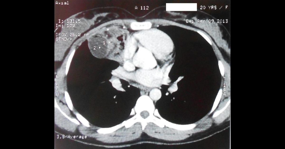
A 20-year old Indian female with a weight of 66 Kg height of 157 cm, presented with chest pain mainly on the right side with occasional nonproductive cough of six months duration. There was no history of smoking, tobacco addiction, tuberculosis, or any significant chest disease. The chest X-ray showed relatively homogeneous lobulated opacity with air lucency within it in anterior mediastinum obscuring right cardiac border (Figure-1).
Contrast-enhanced computed tomography (CECT) of the thorax showed well defined non-enhancing soft tissue density lesion of 97x52x60 mm in the anterior mediastinum with the presence of multiple small well defined cystic lesions showing fat density as well as multiple calcifications within it (Figure-2). Bronchoscopy revealed no intraluminal growth and no bronchial compression at any level. Bronchoalveolar lavage smears were negative for malignant cells.…





