
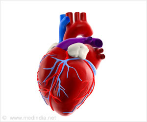
Heart experts, including one of an Indian origin, have successfully integrated two common imaging techniques to produce the first 3D anatomic model of a patient's heart. This is the first time the integration of computed tomography (CT) and 3D transesophageal echocardiography (3DTEE) has successfully been used for printing a hybrid 3D model of heart. The study also opens the way for these techniques to be used in combination with a third tool magnetic resonance imaging (MRI).
"This is a huge leap for individualised medicine in cardiology and congenital heart disease. The technology could be beneficial to cardiologists and surgeons," said Dr Joseph Vettukattil from Helen DeVos Children's Hospital in Michigan. The model will promote better diagnostic capability and improved interventional and surgical planning, which will help determine whether a condition can be treated via transcatheter…