
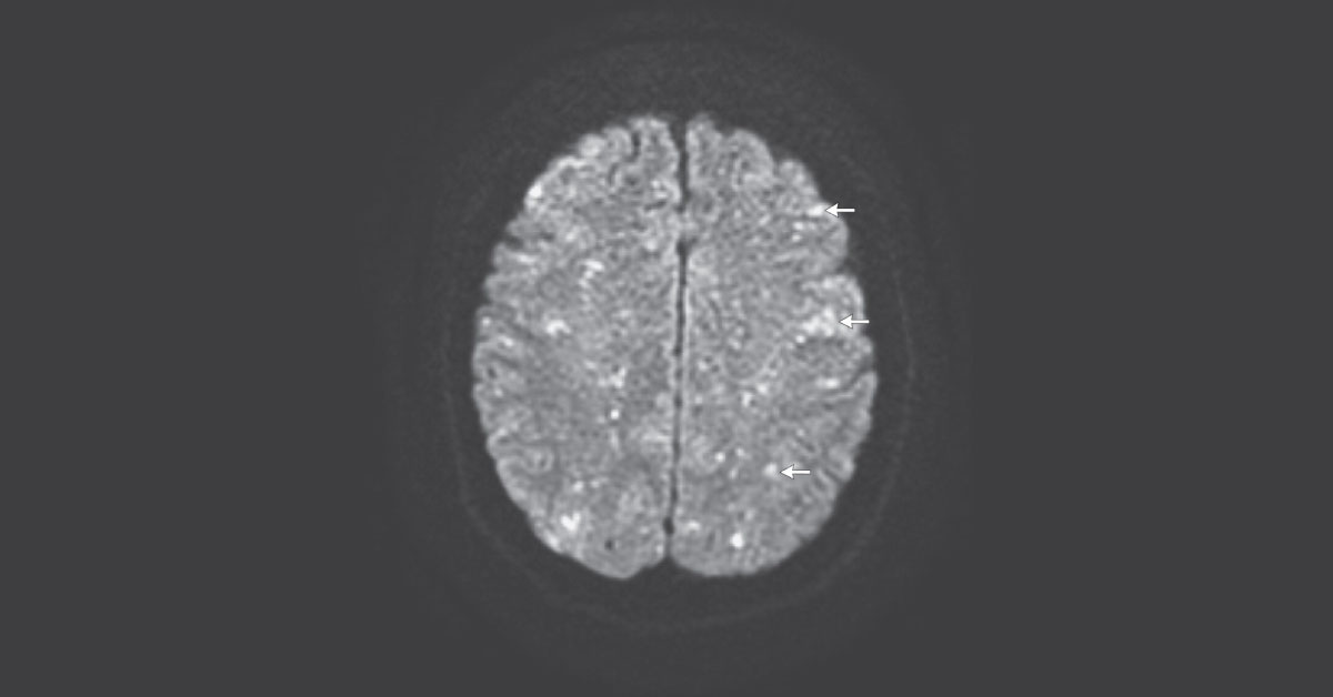
A 56-year old underwent elective hip replacement after right-hip fracture. She remained somnolent a few hours after surgery and required intubation. She had rowing eye movements and decerebrate posturing. Diagnostic Imaging T2-weighted MR images of the brain showed numerous pinpoint, hyperintense foci in both the gray and the white matter of the cerebral and cerebellar hemispheres, the thalami, and the caudate heads.
Diffusion weighted images indicated innumerable punctate foci of restricted diffusion, producing a “starfield” appearance (as indicated by arrows). Identify this neurological condition from attached DWI MR image? *This case is from Docplexus editorial team for educative purpose only Source: NEJM





