
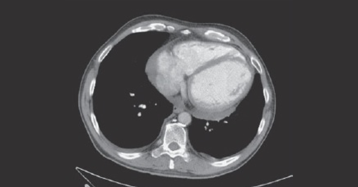
A 66-year-old woman is admitted to ED for sudden onset of chest pain 1 hour earlier. She has a long history (11 years) of hypertension and has been consuming amlodipine. Examination ECG showed evidence of left ventricular hypertrophy. Troponin was not raised but D-dimer was moderately elevated Chest radiograph and CT pulmonary angiogram (CTPA) was performed.
(Please refer to attached respective images). No pulmonary embolism or focal lung abnormality was observed. What abnormalities do the chest radiograph and CTPA images demonstrate? *This case is from Docplexus editorial team for educative purpose only Source: 100 Cases In Radiology