
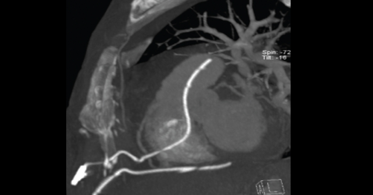
A 43-year-old woman was presented in ED with progressive dyspnea. Case History The woman was diagnosed with malignant pericardial effusion and PET scan was suggestive of primary malignant tumor in the left upper lobe of the lung. The woman also had mediastinal lymphadenopathy. Examination & Procedure Chest radiograph indicated substantial enlargement of the cardiac silhouette. Echocardiography indicated to pre-tamponade. The patient underwent pericardial drainage and 2000 Ml of hemorrhagic fluid was evacuated.
After 24 h, the patient had substantial loss of red blood cells and became hypotensive. Control echocardiogram still showed signs of pre-tamponade. Re-drainage of the pericardium was started and a preoperative CT angiography scan was obtained (attached images). What must have gone wrong during the first pericardial drainage? *This case is from Docplexus editorial team for…