
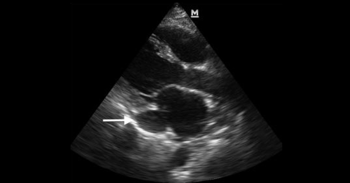
A 55-year-old female was presented to the emergency department with chest pain, dyspnea, and lightheadedness. The symptoms were seen since past one week. She had a history of congestive heart failure, breast cancer, asthma, and right upper extremity port-related deep venous thrombosis. She also endorsed worsening dyspnea on exertion and orthopnea. She denied a fever or productive cough. On examination, she was mildly hypotensive and her electrocardiogram demonstrated low voltage.
A cardiac ultrasound was performed for further diagnosis of her condition (image attached). What does the ultrasound image show? *This patient case is from Docplexus Editorial Team for educative purpose only. Source: American Journal of Emergency Medicine Correct answer has been posted in the comments section. ( Click here ).