
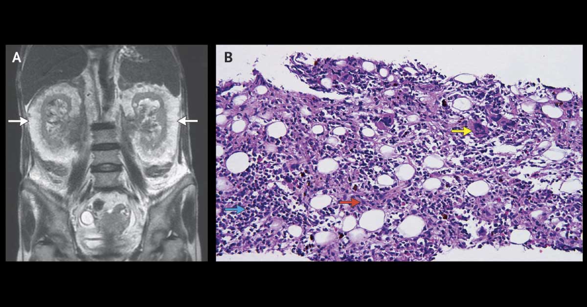
A 69-year-old woman with a history of CKD and myelofibrosis was presented in ED with a 2-day history of dyspnea and general weakness. Laboratory Examination Hemoglobin : 7.2 g/dL (reference range, 11.1 to 15.0g/dL) Hyperkalemia Serum creatinine level: 6.7 mg/dL (0.4- 1.1 mg/dL) Diagnostic Imaging Ultrasonography revealed hyperechoic structures around both kidneys. MRI performed without contrast administration indicated splenomegaly, a normal-appearing liver, ascites, and rim-like lesions on both sides of the perirenal space (Panel A, arrows). Histopathology A biopsy specimen was taken from the left perirenal lesion.
It showed erythroid and myeloid precursors (Panel B, blue and red arrows, respectively) and megakaryocytes (Panel B, yellow arrow), with focal hemosiderin deposition. What is the most likely diagnosis in this case? *This case is from Docplexus editorial team for educative…