
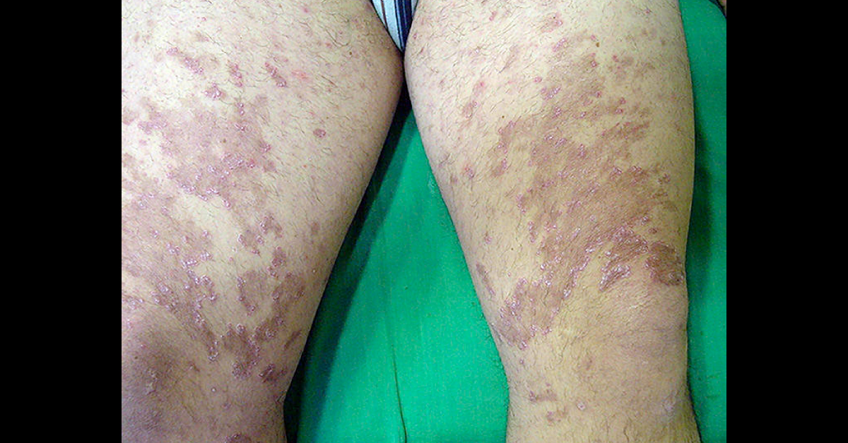
Medical History: A 16-year-old boy with Down syndrome presented with large, asymptomatic, plaques on both lower limbs that had first appeared several months earlier. Physical Examination: Physical examination revealed papules coalescing into annular plaques with hyperkeratotic borders and central depression on the anterior aspect of both thighs. The lesions left residual hyperpigmentation (image attached). No other relevant lesions were found.
Histologic Findings: Analysis of a skin biopsy specimen from the edge of a plaque revealed an acanthotic epidermis with hyperkeratosis, without the presence of intraepidermal neutrophils. Transepidermal elimination of dermal basophilic material was also observed (image attached); orcein staining revealed that the material consisted of elastic fibers (Image attached). What is the most likely diagnosis for this condition? *This image based case is…