A 27-year-old female patient, presented with left hemiparesis. Brain MRI showed multiple cystic ring-enhancing lesions involving the cerebrum and cerebellum. She has received anti-tuberculous treatment for a recent diagnosis of pulmonary tuberculosis, however, the brain lesions showed no response. Investigations revealed no other medical conditions.
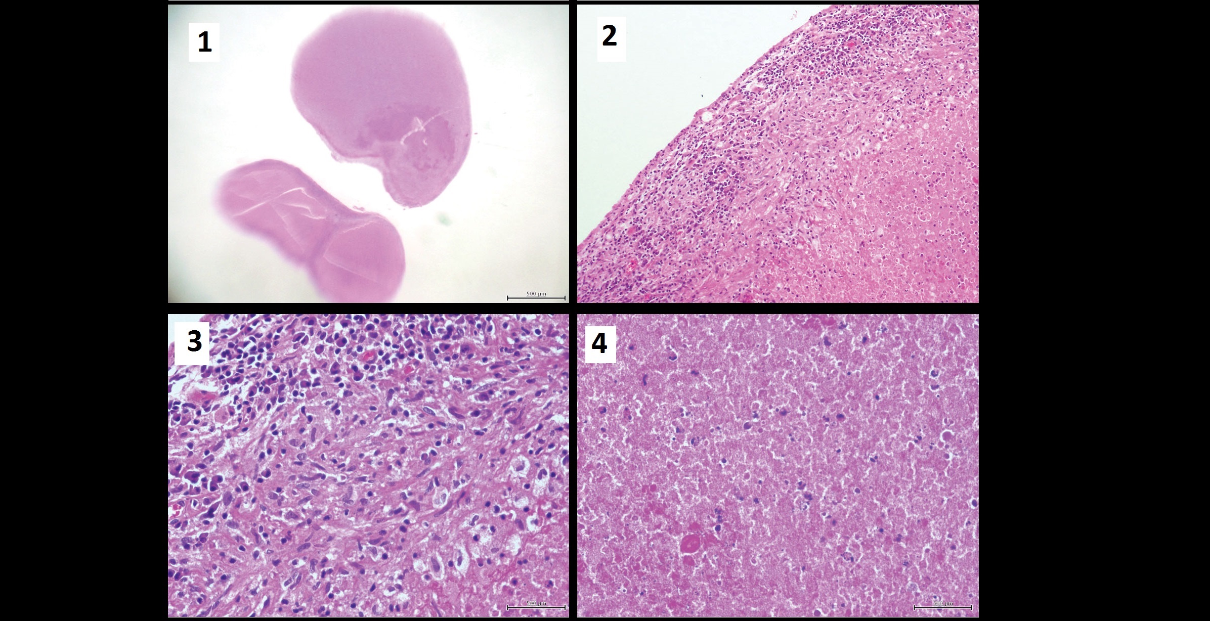
She underwent craniotomy and one of the lesions was excised from the left parietal lobe. It consisted of an oval white to gray soft tissue mass with a solid white cut surface measuring 1.2x0.8x0.9cm. Microscopic examination Microscopic examination revealed a well-defined lesion (Figure-1 H&E X4) that consisted of well-defined chronic granulomatous inflammation (Figures-2 and 3 H&E X10, X20) located at the periphery surrounding a large center of necrosis (Figure-4, H&E X40). *This case is from Docplexus editorial team for educative purpose…