
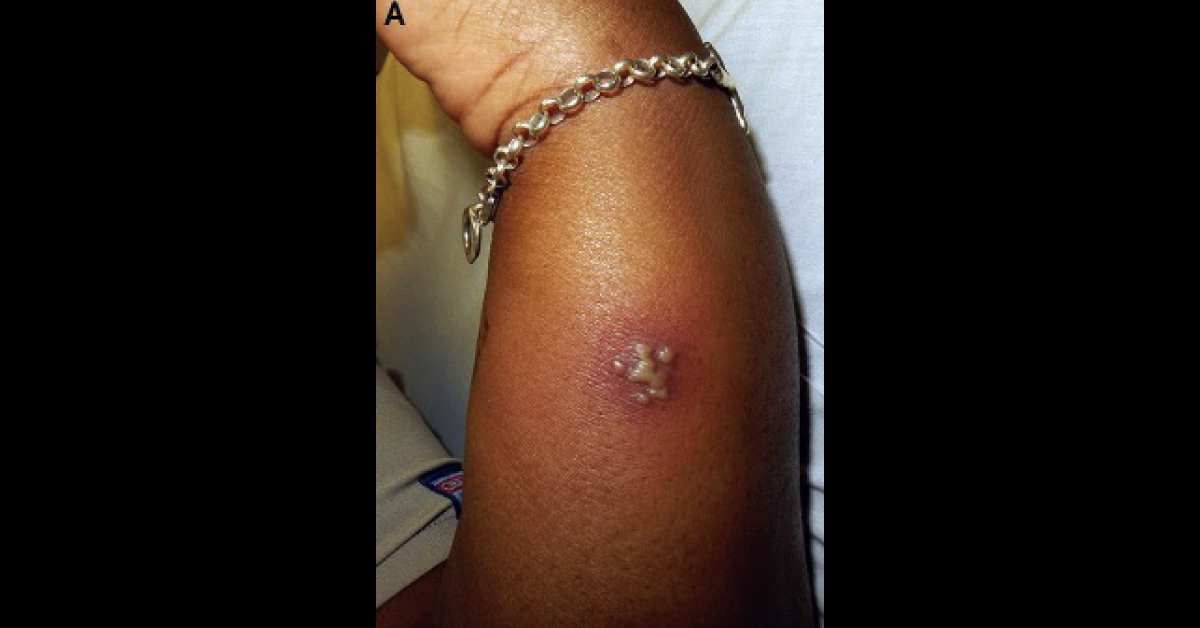
A 27-year-old woman with a history of genital herpes presented to the ED with painful, burning lesions on her left arm. Examination Grouped, coalescing pustules that had scalloped borders on a red (erythematous) base were seen on the left arm (Image A). The pustules were tender to palpation. One of the pustules was unroofed and was smeared onto a glass slide, and stained for Wright-Giemsa (Tzanck smear). Microscopic examination revealed numerous multinucleated giant cells with molded nuclei and marginated chromatin (Image B).
Which infection could this be? How would you manage this patient? *This case is from Docplexus editorial team for educative purpose only. Source: The New England Journal Of Medicine





