
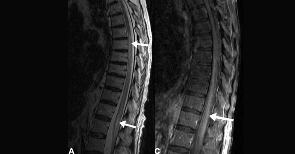
Case A 60-year-old female presented with sudden onset of paraplegia. In the past, she has had a surgery for an abdominal aortic aneurysm. Diagnostic MRI T1 and T2- weighted longitudinal and axial scans were performed and following observations were noted: T2-weighted images of the thoracolumbar region indicated hyperintense signal in the cord extending from T5 to the level of conus medullaris (as indicated in the figure) There was no widening or swelling of the spinal cord. Post-contrast sagittal image indicated a hyperintense signal in the thoracolumbar region (refer figure).
Diagnostic CT Contrast enhanced axial CT abdominal scans indicated large infrarenal abdominal aortic aneurysm with a thrombus. However aortic dissection was not present. Axial CT scan also indicated that the left kidney was small and atrophic in size. Left kidney also showed some parenchymal enhancement in the…