
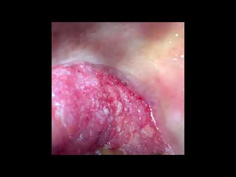
This patient had this slow-growing lesion in the roof of the mouth for the last few months. He had occasional bleeding from the region and also had a pin on and off. She had sought some earlier treatment once before. Recently when he visited the dentist, he was referred to our Hospital for further treatment. What was the lesion? The lesion was an ulcerative and proliferators type of pretty vascular lesion that is around 2x 3 Cm in dimension and lies between the upper first premolar and the second molar on the right upper arch.
As the lesion looked suspicious, a cone beam CT scan was ordered to look for bony involvement. Cbct showed some bony resorption and so we planned an incisional or a wedge biopsy. How was the surgery done? The surgery was done in the operating theatre under local anaesthesia. Therefore this was a daycare surgery. So these types of surgeries are usually performed…





