
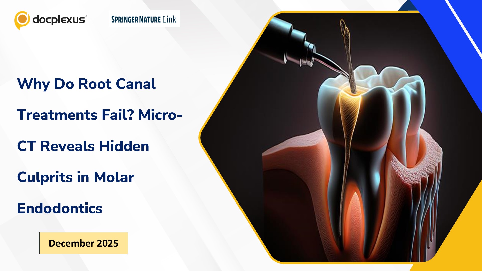
A recent micro-CT–based analysis of failed endodontically treated first molars uncovers critical anatomical and technical shortcomings that often go unnoticed during routine practice. The study identified high rates of untreated canals —particularly a second mesiobuccal canal in maxillary molars (70.6%) and a middle mesial canal in mandibular molars (50%) —along with substantial apical filling voids and a notable prevalence of short obturations. These microstructural deficiencies underpin persistent infection and treatment failure, emphasizing the need for meticulous canal identification, enhanced imaging, and improved obturation techniques to optimize long-term success in molar endodontics.
To read more ; Click here Are you confident you're locating and adequately filling every canal in complex molars? ##Reference## Keskin NB, Odabaşı Tezer E, Uslu G, Tayman AM, Gündoğar M, Kemeç UC,…





