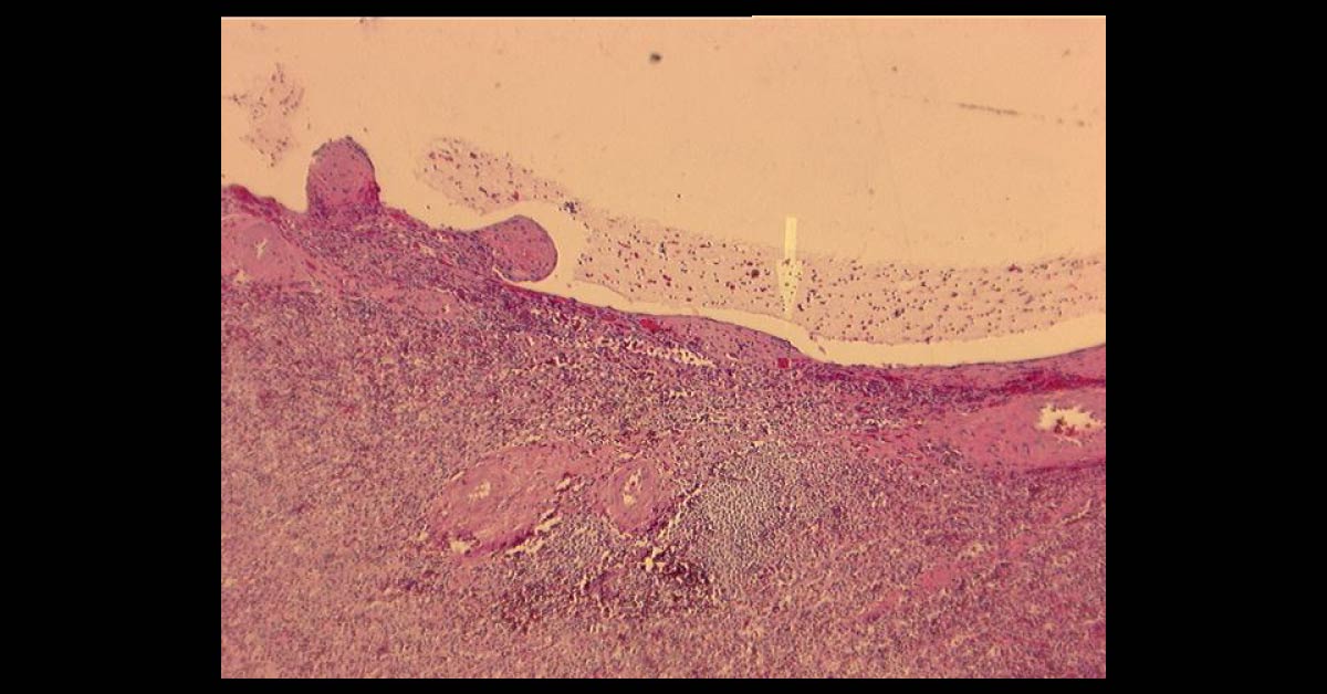
A 60-year-old man with a previous history of thoracic aortic aneurysm was currently presented to the doctor with an abdominal aortic aneurysm and liver and kidney masses. He was admitted for liver and kidney transplant. After the surgery certain microscopic examinations were done which showed: Sections from the liver (images attached) with extensive cyst formation affecting more than 90% of the liver parenchyma were obtained The limited amount of liver tissue showed a variety of changes varying from atrophy to hemorrhage The epithelium lined cysts were seen in the kidney Some cysts had ruptured and lead to hemorrhagic necrosis, calcification and fibrosis.
Focal cholesterol clefts and foreign body giant cells were seen The spleen showed an expanded red pulp and multiple cystic spaces lined by flattened cells What is the diagnosis? *This patient case is from Docplexus Editorial Team for…