
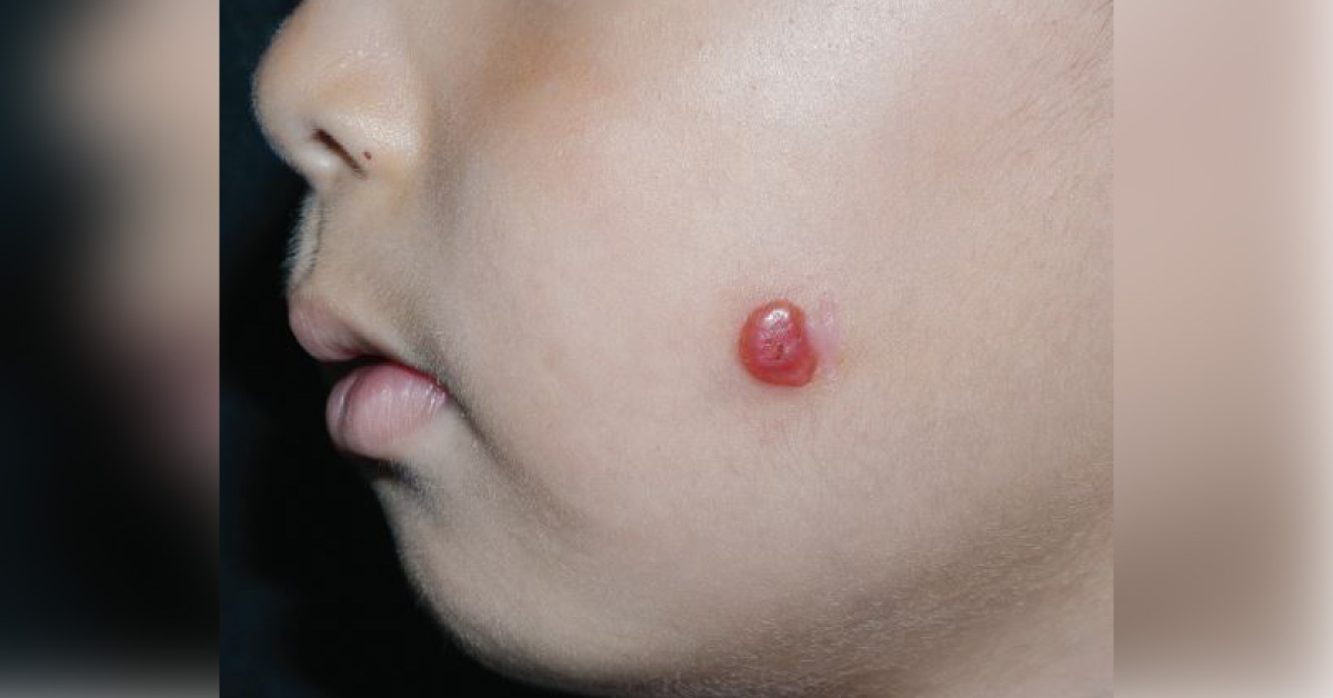
A 4-year-old boy presented to the clinic with a pink nodule on his left cheek. The lesion started as a hyperpigmented macule 3 years ago and since then had been gradually increasing in size and developing into a pink nodule. The lesion was pruritic at times and sometimes bled with minor trauma. The boy was otherwise healthy, with no medical history of trauma or exposure to radiation. On physical examination, there was a 1-cm well-circumscribed, symmetrical, pinkish, dome-shaped nodule with central erosions on the left cheek (See attached figure).
Hair, nails, and mucosae were of normal appearance, and the remainder of the examination was unremarkable. A biopsy was performed and the image of histopathological examination of the lesion is attached. What does the histopathology show? What is the possible diagnosis? *This patient case is from Docplexus Editorial Team for educative purpose…





