
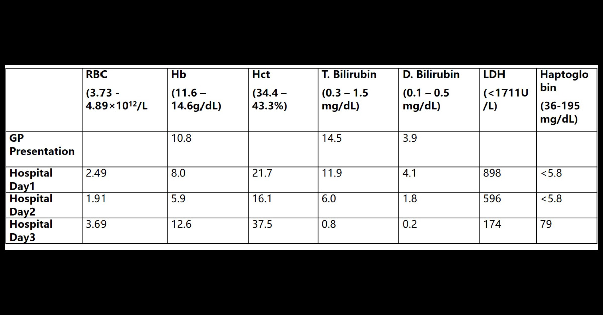
A woman is her late 30's with a history of recurrent scalp lesion. She presented to her primary care physician with a 3-day history of inflamed scalp lesion and an enlarged lymph node. Physical examination: Erythematous skin breakdown at the crown to the right of the midline, but no drainage expressible. A large, tender right posterior auricular lymph node. She was treated with an antibiotic combination containing a sulfonamide for suspected scalp cellulitis. One week after her initial presentation, she developed a fever, jaundice, emesis and coca cola-colored urine.
She was admitted to another hospital for further evaluation. On admission, she was hemodynamically stable. Physical examination revealed scleral icterus. Relevant laboratory results are shown below. Laboratory Results – attached as an image Blood Bank Test Results upon Admission Direct antiglobulin test polyspecific: 3+ …