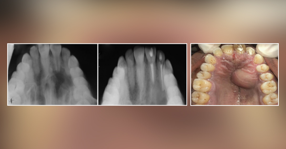
A 30-year-old male reported with a painless, localized swelling in the maxillary right anterior palatal region. A history of trauma was present six years back. Initially the swelling was pea-sized, which gradually increased to the size of an almond. On intraoral examination, tooth no 11 was discolored with a diffuse palatal swelling extending from 11 to 15, about 2 cm × 1.5 cm in size. Palpation revealed the swelling was non-tender, fluctuant, and non-pulsatile with no mobility of the involved teeth.
OPG and intra oral periapical radiograph revealed: A well-defined radiolucency with corticated margins Negative EPT (Electric pulp test) w.r.t. 11 and 12 Loss of lamina dura, external root resorption with 11 Fine-needle aspiration cytology under local anesthesia exhibited a yellow color fluid. Histopathology showed numerous acute and chronic inflammatory cells. The image is attached for…