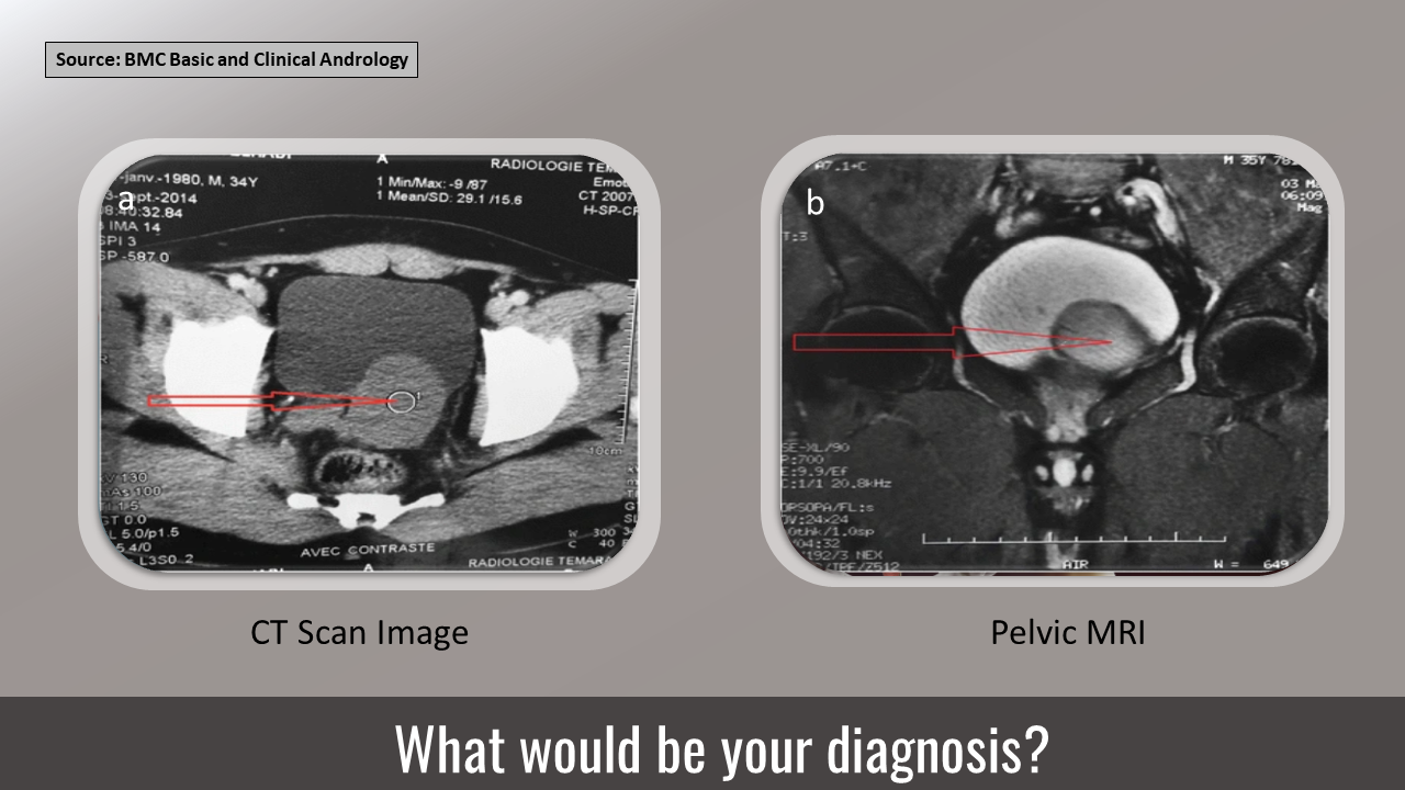
Case presentation A 35-year-old male was hospitalized for subocclusion. He also complained of painful urination. Examination Ultrasound showed a retrovesical liquid mass. CT showed a polylobed contoured liquid mass (Cover image a) The mass measured 80x60 mm, associated with a single right kidney. Pelvic MRI – left-lateralized retrovesical coarsely rounded mass with thin walls measuring 56 mm x 54 mm x 54 mm (Cover image b and Figure 1) No lymphadenopathy No iliac or pelvic effusion Figure 1. Sagittal section of the cyst.
Figure source: Basic Clinical Andrology Figure 2. Image of the laparoscopic surgery of the cyst. Figure source: Basic Clinical Andrology Treatment Cysts were removed surgically (Figure 2). No postoperative complications. Source : Basic Clinical Andrology ##Disclaimer## The content provided on Docplexus is intended for educational purposes only. While we strive to…