
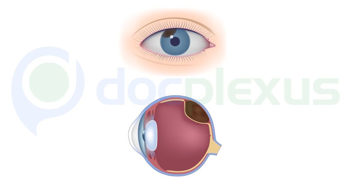
Choroidal melanomas are one of the commonest intraocular tumors can be benign malignant pigmented or non-pigmented 70 % are choroid 20 % involve ciliary body and 10 % iris. They are difficult to diagnose due to their atypical presentation, however in most of situations are seen as solid or exudative retinal detachments on ophthalmoscopy both direct and indirect. B scan ocular ultrasound other diagnostic modalities are CT scan, brain MRI and FF angiography their atypical manifestations. Melanoma to lens will present as blurred vision paracentral scotoma progressive and painless visual field impairment.
If melanoma involves angle of filtration the presentation is Angle closure glaucoma. Angle closure glaucoma ocular hypertension or even chronic uveitis may involve vitreous. It present as vitreous floaters sometimes severe ocular pain may be only presentation very rarely severe…