
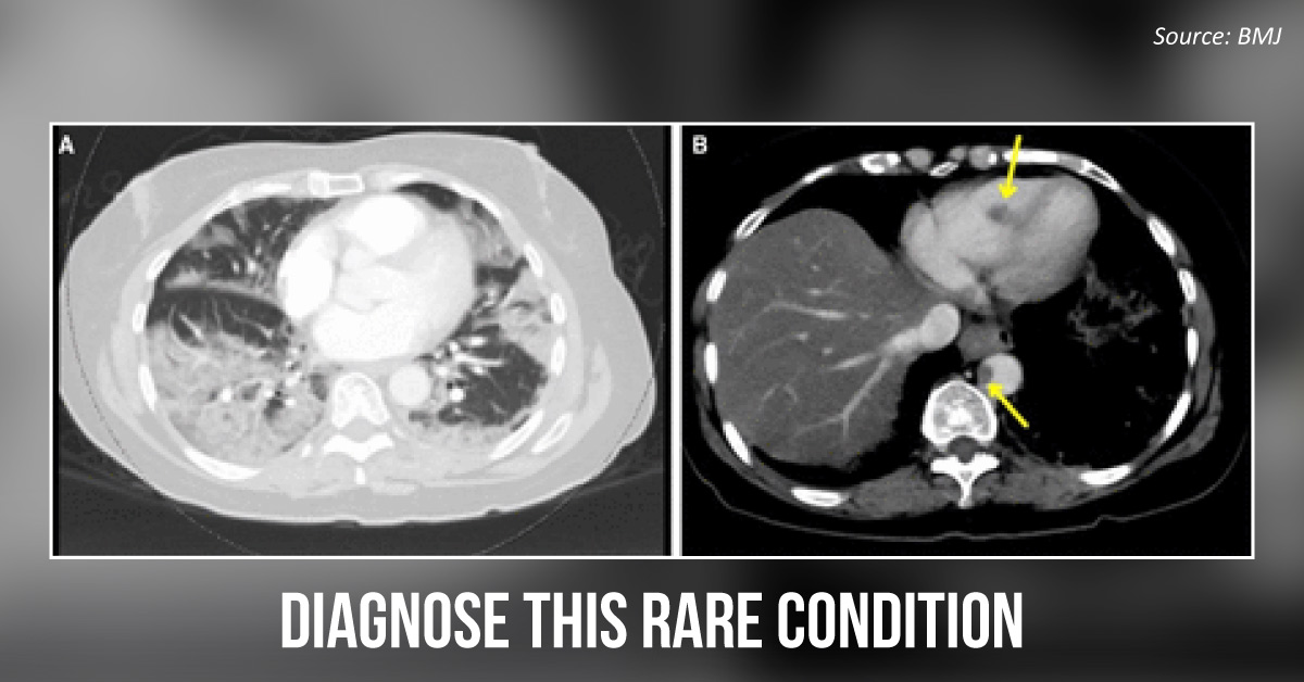
Case presentation: A 61-year-old woman presented in the ER with acute onset 10/10, non-radiating midabdominal pain, and dry cough. Medical history: Type 2 diabetes mellitus, hypertension, hyperlipidemia, bipolar disorder, and gastroesophageal reflux disease Laboratory investigations: RT-PCR test via nasopharyngeal swab was positive for COVID-19. ECG revealed sinus tachycardia. Radiological investigations: CT-pulmonary angiogram revealed bilateral peripheral ground-glass opacities with intralobular septal lines and areas of dense consolidation.
( Cover image A) Hypoattenuating focus area within the right ventricular apex and thoracic aorta was observed. (Cover image B) Emergency point-of-care ultrasound (POCUS) showed an echogenic mass measuring 19×14 mm in the RV cavity. Based on the radiological images and the laboratory investigations, what is your diagnosis for this patient? A.…