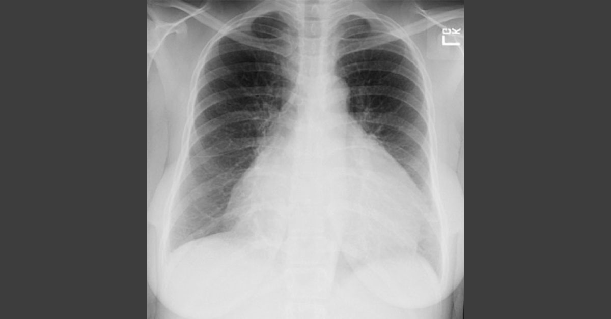
A 50-year-old female with chest pain. Image 1: Frontal chest radiograph Image 2: A single axial, contrast-enhanced CT scan of the chest *This image based case is from Docplexus editorial team for educative purpose only Source: Learning radiology
Deduce The Conclusion From The Image

Dr. Kapil Patil
Docplexus Editorial Team