
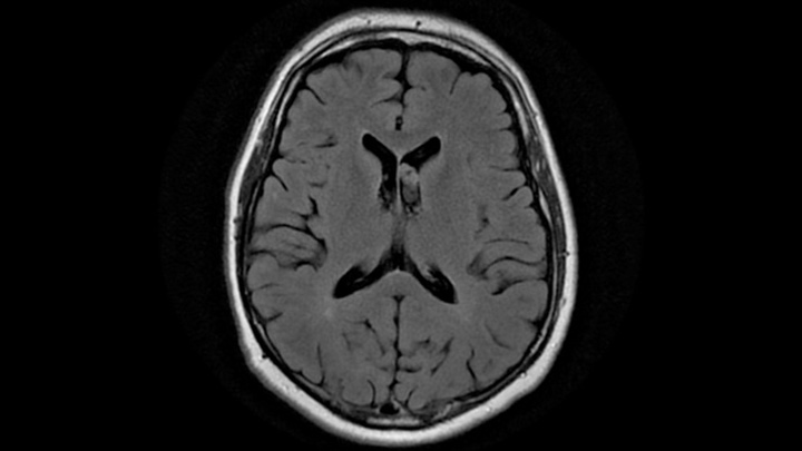
Case Male 55-year-old patient. No patient history is available. There are no changes in the lesion for last 5 years. Attached are the images of axial T1-weighted and T2-weighted sequence.
Following observations were made from MRI images: Intraventricular lesion in the frontal horn, however, no change in ventricular configuration No intrinsic T1 hyperintensity was detected Lesion was mildly hyperintense on T2-weighted sequence (FLAIR) No parenchymal lesion What condition possibly this be? *This case is from Docplexus editorial team for educative purpose only. Source: Radiopaedia





