
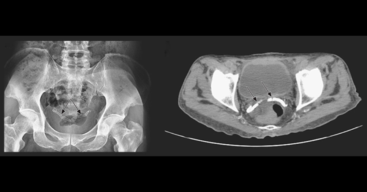
A 60-year old man underwent abdominal and pelvic region radiographic imaging as a part of the evaluation for nephrolithiasis. This patient has advanced diabetic nephropathy and secondary hyperthyroidism. Panel A: Radiograph; Panel B: CT image, without contrast Radiographic findings showed no hydronephrosis or radiopaque kidney stones. But the axial section of a CT image of the pelvis showed bright regions in the vas deference.
Identify this condition in this diabetic patient. * This case is from Docplexus editorial team for educative purpose only. Source: NEJM Stay Connected, the answer will be posted in the next 48h!





