
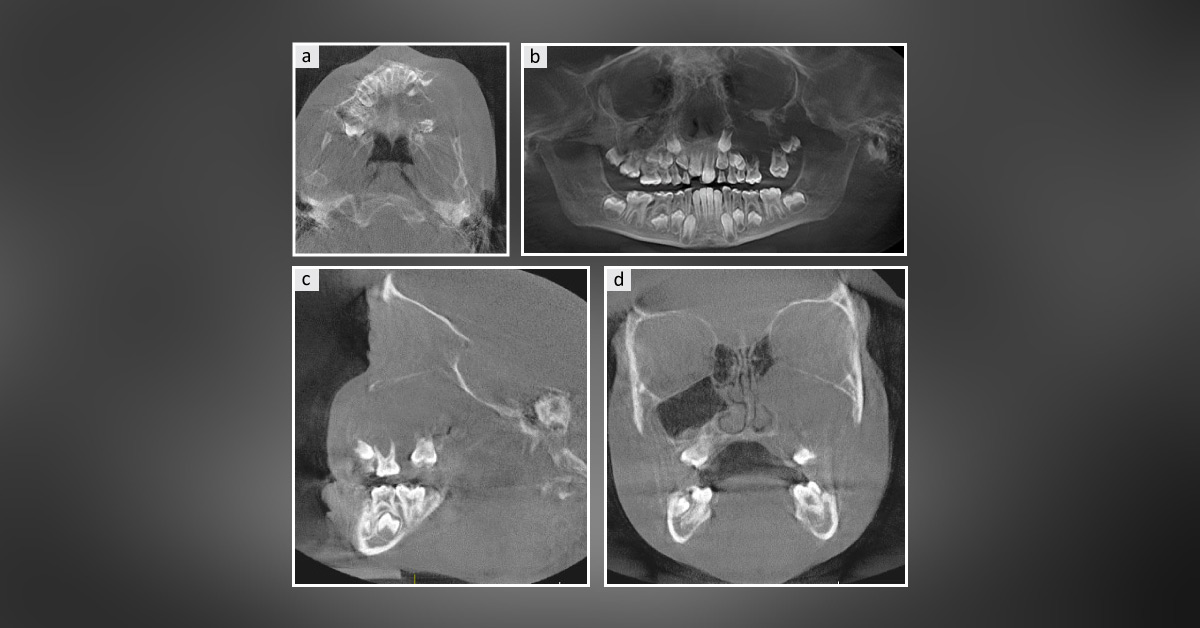
Case presentation A 7-year-old female patient was referred to a dentist to get swelling on the left cheek examined that was noticed recently. Subsequently, she was referred to an oral surgeon to get a large vestibular swelling and cyst-like lesion involving the left maxilla evaluated. The swelling was not painful, and there was no neural dysfunction. The past medical history was not significant. Examination Cone beam computed tomography (CBCT) revealed a large radiolucent lesion at the left posterior maxilla filling the left maxillary sinus (Figures a, b,c, and d).
Intraoral examination showed the fullness of the maxillary vestibule and loss of vestibular depth. It was asymptomatic and firm upon palpation. Histologic examination revealed multiple pieces of soft tissue composed of loose and myxoid connective tissue. The immunohistochemistry stain was negative with S100, which rules out…



