
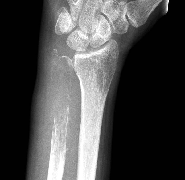
A 72-year-old man visited the surgical outpatient clinic with a spontaneous fracture of his left wrist. Physical examination suggested an additional fracture of his right wrist. Conventional radiograph showed extensive osteolytic lesions with fractures of both distal ulnae (figure). He reported no recent weight loss, night sweats, prostatic problems, or deteriorating performance status. There was no lymphadenopathy on physical examination. Positron emission tomography showed no primary tumour in the abdomen or thorax and no other skeletal lesions, nor was there any suspicion of prostate carcinoma.
What are the next course of action that will lead to the diagnosis? What can be the probable diagnosis looking at the image? Source: The Lancet. Detailed citation of source will be with the explanation . *This case is from Docplexus Editorial Team for educative purpose only.





