
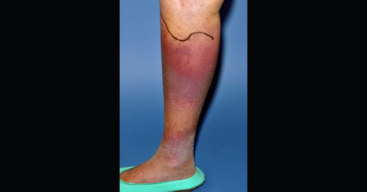
A 79-year-old man presented with the 4-weeks history of progressive discoloration of both lower legs. Patient history His GP had given two courses of antibiotics for possible cellulitis but there was no clinical improvement. He was systemically well and had no history of fever. He denied any trauma to the legs and had no dermatological history. He was taking no regular medication. The man lived alone and was fully independent. His diet lacked fruit and vegetables.
Exam Normal pulse, blood pressure, and oxygen saturations and no fever. Chest and abdominal examination were unremarkable. There was widespread bruising (ecchymosis) of the lower legs with “woody” edema (image 1). Inspection of the legs more proximally revealed perifollicular hemorrhage and “corkscrew” hairs (image 2, fig 3). The right leg appeared larger than the left but the calf was soft and non-tender. Labs A coagulation…