docplexus
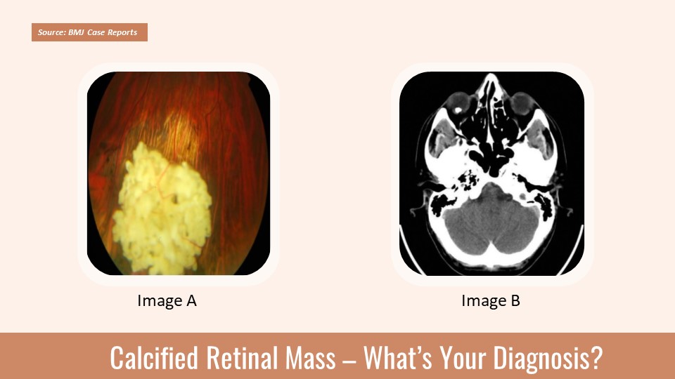
Case presentation A 28-year-old healthy male reported to an ophthalmology emergency department with a six-month history of photopsia of his right eye. Examination Best corrected distance visual acuity of 1.0 (right eye)/1.2 (left eye) Unremarkable anterior segment examination at the slit lamp Fundoscopy Unremarkable posterior pole in the right eye (Figure 1) Figure 1. Posterior pole retinography (right eye) Inferior periphery retinography Presence of a white voluminous subretinal lesion (Cover image A) The lesion was surrounded by pigment epithelium atrophy (Cover image A).
Presence of hypertrophy in the inferior quadrants Ocular ultrasound The presence of a mass with high reflectivity, having an apical height of 4 mm and 6 mm at its largest basal dimension A cranial CT scan confirmed the tumor calcification (Cover image B). Source: BMJ Case reports ##Disclaimer## The content provided…