
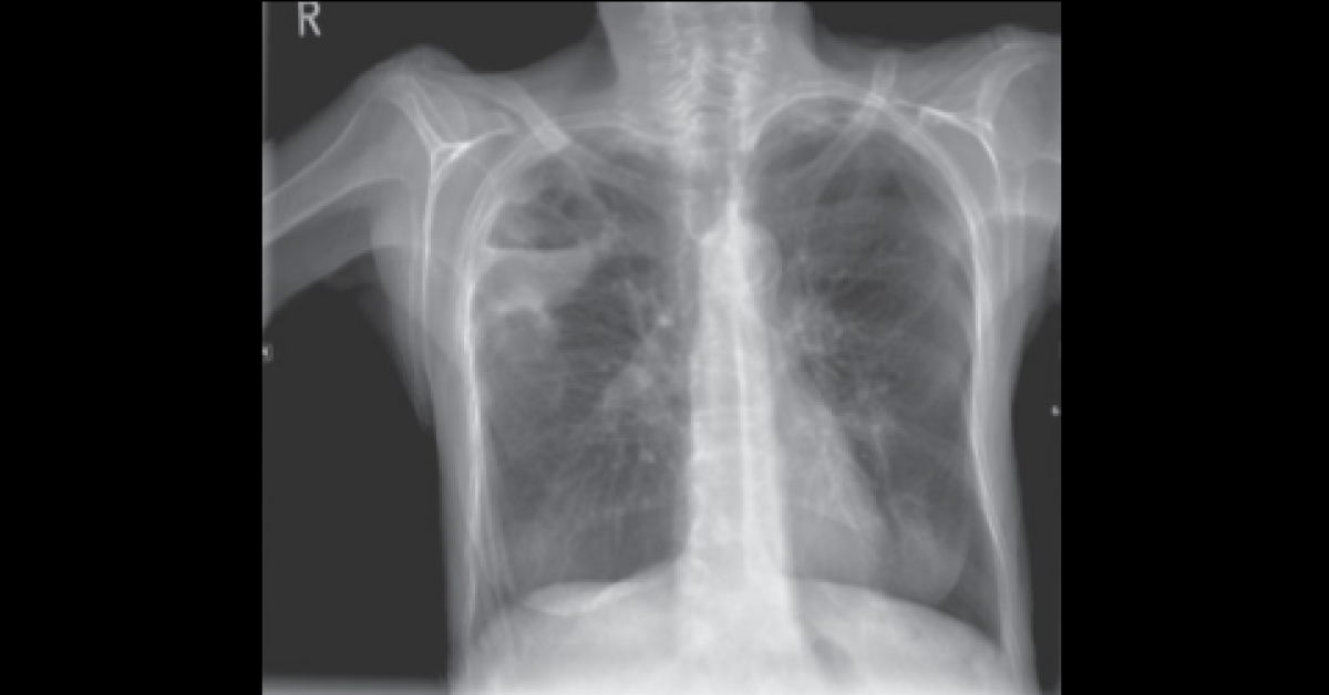
A 66-year-old woman was referred to the emergency department by her GP with a 2-week history of worsening dyspnea, cough and fever. She had a past medical history of type 2 diabetes and high blood pressure treated with appropriate medication. The GP had treated her for a few days with antibiotics initially with little effect.
On Examination: Temperature - 39.3°C Pulse - 104/minute Oxygen saturation - 89 % breathing air and there appears to be a right ventricular heave RS - coarse lung sounds in the right upper zone posteriorly P/A- soft and non-tender What does the chest radiograph reveal? *This case is from Docplexus editorial team for educative purpose only Source: 100 cases in radiology The answer is posted in the comments section. Click here