
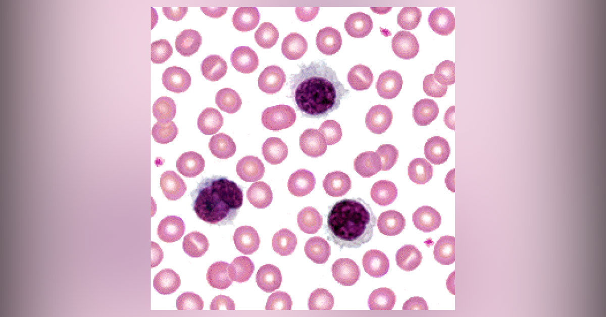
A 51-year-old male was examined by his physician for a routine preoperative exam before dental surgery. The physician finds low hemoglobin and a large left upper quadrant mass. Examination Splenomegaly was noted (which was subsequently confirmed by CT. Blood smear was performed. RBC morphology looked normal. There were some abnormal cells with round or indented nuclei with coarse chromatin patterns.
Some of the cells also had irregular borders and numerous projections (refer attached image). Laboratory investigation was follows: RBC : 3.36 x 10 12 /L HGB: 10.9 g/dL HCT: 31.2 % MCV : 92.8 fL MCH: 32.4 pg MCHC : 34.9 g/dL WBC: 9.3 x 10 9 /L. What is the most likely diagnosis? *This case is from Docplexus editorial team for educative purpose only. Source: Department of Hematology, University Of Minnesota. Stay Connected, the answer will be posted in the next 48h!





