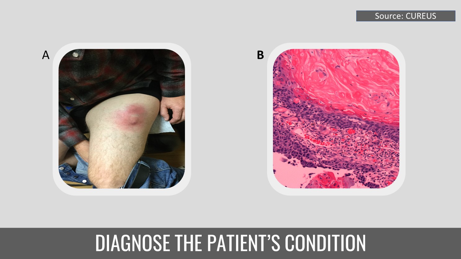
Case presentation A 51-year-old Caucasian man with no history of skin cancer presented with a slow-growing lump on his left thigh that had been there for six months. On physical examination, a 6 cm x 6 cm erythematous subcutaneous lump with surrounding inflammation was found on the lateral portion of his left proximal thigh (Cover image A). The patient was prescribed oral antibiotics and told to apply warm compresses after the lesion was initially suspected to be an infectious cyst.
Laboratory tests Biopsy was performed post two weeks, and pathology revealed calcified homogeneous keratin material with ghost cells, distinct multinucleated giant cells, and lymphocytes. (Cover image B) Due to severe calcification, an initial conservative excision was performed, following which the patient was sent for additional surgical therapy. The pathological findings revealed regular tumor lobules…