
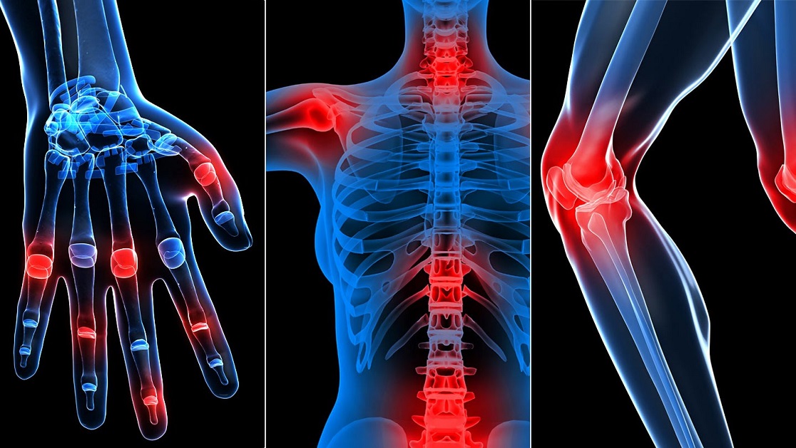
The first-line imaging technique for diagnosis inflammation in musculoskeletal organs in rheumatoid arthritis (RA) is planar X-ray examination, which was for many years the first and the only single tool for RA diagnostics and response evaluation. But today, in the era of more aggressive RA treatment, ultrasound examination (US) and magnetic resonance imaging (MRI) are also frequently used. This article focuses on the different imaging techniques used in rheumatology. In contrast to X-ray, MRI, CT and US allows assessing both the soft tissue and the bone marrow affected by inflammation that precedes the development of destructive lesions observed by X-Ray of CT.
The advantages of MRI It allows assessment of bone marrow inflammatory lesions and spinal lesions. It has higher specificity due to the ability to use multiple sequences and i.v. contrast-enhanced scans. MRI scans of individual…