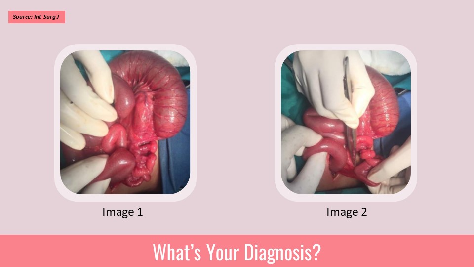
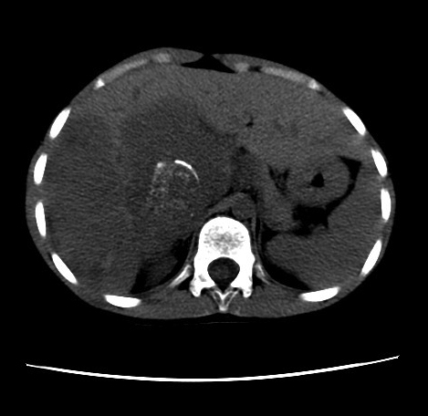
Case: A 9-year-old female was found to have liver lesions on a computed tomography (CT) exam which was performed due to mild, ongoing abdominal pain associated with nausea and vomiting. Imaging Findings: One noncontrast-enhanced and three contrast-enhanced axial images of the liver through the abdomen demonstrate a large, multilobulated lesion that replaces the majority of the left lobe of the liver. The mass is of heterogeneous density, though predominantly hypodense with scattered foci of thin linear, discontinuous peripheral enhancement.
What is the likely diagnosis for this case? Source: Appliedradiology *This case is from Docplexus Editorial Team for eductaive purposes only