docplexus
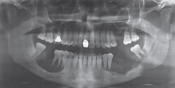
A 36-year-old man presented with a 1-year history of oral pain and loose teeth; four teeth had been lost spontaneously within the 6 months before presentation. Increased thirst and urination also developed within 4 months before presentation. Blood electrolyte levels revealed hypernatremia and dehydration. A panoramic radiograph showed two radiolucent scooped-out lesions of the mandible with “floating tooth” images.
(Image attached) Biopsy of the mandibular lesion on the left side revealed sheets of inflammatory-cell proliferation and S100+ CD1a+ Langerhans’ cells. Magnetic resonance imaging of the brain was done. (Image attached) Source: NEJM, Detailed source will be cited with the conclusion * This case is from Docplexus Editorial Team for educative purpose only.